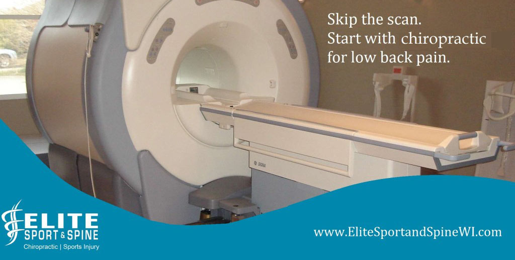
February Research Review: Do MRI Findings Match Musculoskeletal Symptoms?

At Elite Sport & Spine, we pride ourselves on being evidence based chiropractors. Our doctors frequently read the newest research on spinal manipulation, spine care, physical therapy, and rehabilitation. We want to share some of our reading with you, in a manner that is easy to understand.
Our focus this month will be on MRI findings and how they correlate with musculoskeletal symptoms.
1. Abnormal Knee MRI Findings in Asymptomatic Adults
Overview: This study came out on February 3rd, 2020 in the Skeletal Radiology journal. The purpose of the study was to evaluate abnormalities found in the knees of ASYMPTOMATIC individuals. These individuals were considered sedentary (didn’t meet the recommendations for weekly physical activity), did not have any prior knee injuries, and were over the age of 18. The knees were studied using a 3.0 T MRI and then the results were reviewed by multiple radiologists.
Results
Nearly all knees (227/230; [97%]) of asymptomatic individuals showed abnormalities in at least one of the knee structures on MRI, of varying grades of severity. Meniscus tears: 30%. Articular cartilage abnormalities: 62%. Bone marrow edema: 52%. Tendon abnormalities: 46%. Ligament abnormalities: 38%.
What does this mean? Results like this help to further prove that diagnostic imaging (x-rays, MRI, CT scans) findings are poor predictors of musculoskeletal pain. If imaging was a strong predictor, more of the study participants would have experienced symptoms.
Citation: Horga, L.M., Hirschmann, A.C., Henckel, J. et al. Prevalence of abnormal findings in 230 knees of asymptomatic adults using 3.0 T MRI. Skeletal Radiol (2020).
2. Spinal Degeneration MRI Findings in Asymptomatic Adults
Overview: This is a systematic review study that came out in April of 2015 that looked to estimate the prevalence of radiographic spinal degeneration in asymptomatic patients. In this study, it reviewed articles that used CT scans and MRIs to view the spine. Researchers selected groups by decades, with the starting age being the 20s, all the way up to the 80s. In order for a study to be included, the patients in the study had to have never reported any history of back pain. Researchers included 33 articles in their analysis, which led to 3110 asymptomatic individuals reporting imaging findings.
Results
The prevalence of disk degeneration in asymptomatic individuals increased from 37% of 20-year-old individuals to 96% of 80-year-old individuals. Disk bulge prevalence increased from 30% of those 20 years of age to 84% of those 80 years of age. Disk protrusion prevalence increased from 29% of those 20 years of age to 43% of those 80 years of age. The prevalence of annular fissure increased from 19% of those 20 years of age to 29% of those 80 years of age.
What does this mean? Again, the results of a study like this help further prove that findings on diagnostic imaging (x-rays, MRIs and CTs) are poor predictors of a patient with musculoskeletal pain, especially when it comes to low back pain. As chiropractors, we want to avoid labeling patients with these findings. Mostly because they are common and often a natural part of the aging process. In addition, other research has shown these labels can become a massive barrier to patient improvement.
Citation: Brinjikji, W., et al. “Systematic Literature Review of Imaging Features of Spinal Degeneration in Asymptomatic Populations.” American Journal of Neuroradiology, vol. 36, no. 4, 2014, pp. 811–816., doi:10.3174/ajnr.a4173.
3. Suspected Rotator Cuff Injuries MRI Findings and the Role of Conservative Management
Overview: This was a prospective study that came out in November of 2019. The study looked at how often patients, in this study 51 (which is small to be considered statistically relevant), were having MRI’s of their shoulders done prior to a trial of conservative care. It then assessed the cost of these MRI’s in relation to the cost of the conservative care. In order to be included in the study, the patients had to have shoulder pain NOT related to trauma, minimal to no strength deficits, and suspected rotator cuff tendinopathy. Each patient underwent an MRI and had a trial of conservative care.
Results
Of this cohort, 46 (90.2%) patients did not go on to surgical intervention, whereas 5 (9.8%) patients did, at an average 68.3 days after imaging. These results suggest that over 90.2% of patients (46 of 51) had premature MRI, posing an unnecessary economic burden of $181,619 in advanced imaging charges.
What does this mean? It means, you can probably pump the breaks on getting an MRI on your shoulder if you didn’t have a trauma or if you don’t have major strength deficits. As stated in the studies above, findings on x-rays, MRIs or CT scans are not good predictors for musculoskeletal pain. So, looking for answers through an MRI on why you might be dealing with shoulder pain is probably not the most cost effective, and you might not get the answer you were hoping for. It might make more sense to try conservative care and strength training to help improve your rotator cuff strength and improve your shoulder health.
Citation: Cortes A, Quinlan NJ, Nazal MR, Upadhyaya S, Alpaugh K, Martin SD. A value-based care analysis of magnetic resonance imaging in patients with suspected rotator cuff tendinopathy and the implicated role of conservative management. Journal of shoulder and elbow surgery. 2019 Nov 1;28(11):2153-60.
This blog was written by Dr. Adam Cosson
 262-373-9168


